In cells, many vital processes take place in molecular aggregates, which play an important role in signal processing within the cell. Scientists from the University of Freiburg, Germany, and this department have only now been able to observe and analyse how these aggregates form in living cells for the first time.
Writing in the journal Nature Communications, they show that this process is controlled not by physical forces alone, but also by active biological mechanisms. The experimental protocols and analysis tools have been made available free of charge, enabling further research on small aggregates.
Protein aggregates forming in real time.
If molecules inside a cell were completely randomly distributed, the cell would not be viable. Subdivision into more specialised compartments is necessary for many biochemical processes to take place in a coordinated manner. Some compartments are separated from each other by membranes, but many others are not. Such “membraneless” molecular aggregates fulfil important biological functions because their sizes and numbers are particularly flexible. It is often assumed that they form via the physical process of “liquid–liquid phase separation”.
“These condensates are an important control mechanism in cells because they can accelerate or slow down biochemical processes as needed,” explains ProfessorThorsten Hugel, who is a member of the Cluster of Excellence Centre for Integrative Biological Signalling Studies at the University of Freiburg, and who led the current study together with Professor Aleks Reinhardt from this department.
How condensates help the cell to process biological signals and environmental stimuli is still under-researched, says Hugel. “Research usually focuses on large and static condensates because they are easier to study. But these large condensates are usually only the final stage of a long process. In many respects, small condensates that grow and decay dynamically are much more interesting," he explains. The problem: by construction, they comprise relatively few molecules and are thus too small and fast even for high-resolution microscopy methods to study in living cells.
New method circumvents technical limitations
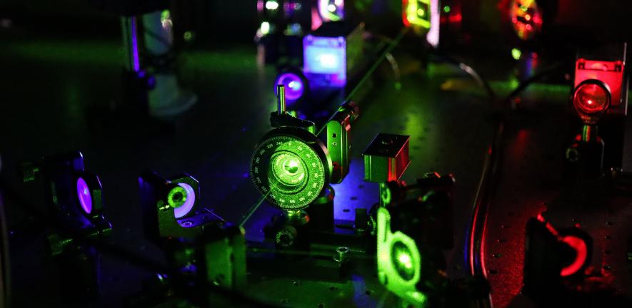
In the current study, the Freiburg and Cambridge scientists describe a way to bypass these technical limitations. In particular, they use conventional high-resolution fluorescence microscopy with a tilted laser, called HILO microscopy, combined with a special experimental procedure and AI-based analysis methods.
The researchers compared the measurements made in living cells with conventional theoretical descriptions of condensate formation. “The results initially surprised us,” says Reinhardt. “For the condensates we studied here, the initial growth still follows familiar physical models, as one might expect. However, once they reach a certain size, their growth suddenly stops.”
Regulated by stress signals
Protein aggregates forming in real time.
In the current study, the researchers investigated aggregates of the protein NELF. These protein aggregates form when a cell is under stress, for example due to heat or when there is an aggregation of other proteins, as happens in dementia and neurodegenerative diseases. “By forming condensates in the cell nucleus, NELF inhibits the expression of genes more effectively,” says co-author Dr Ritwick Sawakar, summarising the protein's natural function. “This inhibition is important for the cell to survive stress.” Sawakar also worked at CIBSS and is currently conducting research at the MRC Toxicology Unit at the University of Cambridge.
The scientists observed that many small NELF condensates also exist in non-stressed cells. “In non-biological systems, we might expect condensates to keep growing once they have reached a critical size. But in living cells, this only appears to be the case when the cell is stressed,” describes Reinhardt. From this, the scientists conclude that NELF condensates are kept small in an active way by the cell until stress signals permit rapid large-scale condensate growth.
Important for signal processing
According to the scientists, although this process may seem complicated at first, it is likely to be essential for the processing of stress signals: “It allows larger condensates to form very quickly and small ones to dissolve when the need arises,” explains Hugel. “This enables the cell to react to stress in time.” Such an efficient response to stress is particularly relevant as we age, as many age-related neurodegenerative diseases are caused by less effective stress responses.
It is generally thought that protein aggregates have many different, fundamental functions for signal processing in cells. The newly developed method enables researches to gain a comprehensive understanding of these functions. Moreover, this also enables research into the role of protein aggregates in misfolding diseases such as dementia and neurodegenerative diseases such as Alzheimer's or Huntington's. In the longer term, a thorough understanding of these mechanisms could therefore help in diagnosing disease and the development of therapies.
Research
Chenyang Lan, Juhyeong Kim, Svenja Ulferts, Fernando Aprile-Garcia, Sophie Weyrauch, Abhinaya
Anandamurugan, Robert Grosse, Ritwick Sawarkar, Aleks Reinhardt, and Thorsten Hugel, Quantitative real-time in-cell imaging reveals heterogeneous clusters of proteins prior to condensation, Nature Communications (15 August 2023)

