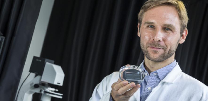
“We are thrilled to have been chosen by the MJFF to develop this new technology, which aims to help clinicians to accurately diagnose Parkinson’s Disease (PD) at a much earlier stage than is currently possible,” said Knowles, who holds the 1920 Chair in Physical Chemistry in this department.
“This is a huge missing piece of the puzzle for understanding Parkinson’s disease. The platform we’re developing will not only enable early diagnosis of the disease but will hopefully also allow researchers to track and monitor disease progression.”
Lewy bodies and alpha-synuclein
In Parkinson’s disease, abnormal deposits of the protein alpha-synuclein form Lewy bodies in the brain, which lead to the characteristic physical and mental symptoms of the disease. Once the aggregates start forming, small strands or fibrils can break off to form new aggregates in a process called secondary nucleation, which was discovered by Knowles and his colleagues ten years ago. The driving forces behind the aggregate formation and seeding of new aggregates are still unknown.
“At the moment, alpha-synuclein-related biomarkers can be picked up, but typically only at roughly the same time as other symptoms become present and detectable,” said Knowles. “There are a lot of indications that the changes in biology occur much earlier, but we don’t have the ability yet to discover what they are. We will be working to identify which of the aggregates is causing the damage, and how and why they are seeding other aggregates.”
New platform
The researchers will use the funding to design a new diagnostic platform based on the group’s pioneering microfluidic technology, which uses microfluidic chips to enable researchers to probe proteins on the scale of a single aggregate.
“It has been very challenging to probe and characterize the pathological seeding behaviour of proteins at the single aggregate level until now through conventional experiments, because the aggregates are very diverse and present only at very low concentrations,” said Knowles. “But we have been able to change this over the past few years by using microfluidics to study aberrant protein behaviour at the single molecule level.”
“Using our custom-developed microfluidic devices we are able to digitally measure the seeding activity of individual aggregates. In digital assay formats, instead of one reaction, you split the sample into millions of smaller compartments, then count. These types of ideas are transforming other areas of biomolecular science through concepts such as digital PCR [used in Covid-19 testing], and it is exciting to bring these ideas to the study of protein aggregation in disease. From the chemistry point of view, it’s a completely new way of doing measurements--it’s a digital solution.”
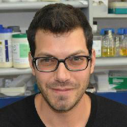 Dr Aviad Levin will help lead the research.
Over the past five years, smaller-scale grants from the MJFF have enabled postdoctoral researcher Aviad Levin to develop a pilot platform. He and other group members have demonstrated the concept can be successfully used to accurately measure and characterize the aggregates and to ultimately find the key bio-markers.
Now Knowles and Levin plan to build a team and scale up the process. “What we’ve done so far is establish the technique,” said Levin. “We have shown we can perform the counting assay, but now we need to automate the process and increase throughput while decreasing background noise. It’s incredibly exciting, and we’ll be able to explore whole new areas.”
A world-wide network
“MJFF have fantastic connections with biobanks and clinicians, and we’re delighted to be part of this world-wide network,” said Knowles. “They have identified the lack of a quantitative biomarker for Parkinson’s disease as one of the top priorities in this space, so we’re very excited to have the chance to work on that. This is a very high priority project for them and for us.”
The work is complementary to the research being done in Professor Sir David Klenerman’s research group, where researchers are using microscopy to identify and characterize individual aggregates. Both the Knowles and Klenerman groups are collaborating actively to push the bounds of measurement science in protein misfolding diseases.