Afilliated Professor
What I do
My research is in the field of molecular imaging, which is the visualisation, characterisation, and measurement of biological processes at the molecular and cellular levels in humans and other living systems. I focus primarily on the nuclear medicine technique of positron emission tomography (PET) - a non-invasive technique based on using positron-emitting molecular probes coupled with sensitive high-resolution scanners to generate quantitative three-dimensional images. This provides an unequalled ability to quantify and assess biological processes in vivo.
Programme collaborations
My programme of research is based at the Molecular Imaging Chemistry Laboratory and the Wolfson Brain Imaging Centre, both situated at the Cambridge Biomedical Campus, and consists of the three main themes outlined below. Central to my research programme, in particular for the first two themes, are collaborations with members of this department including Dr Andreas Bender, Prof Chris Hunter, Prof Sir David Klenerman and Dr Finian Leeper.
Research themes
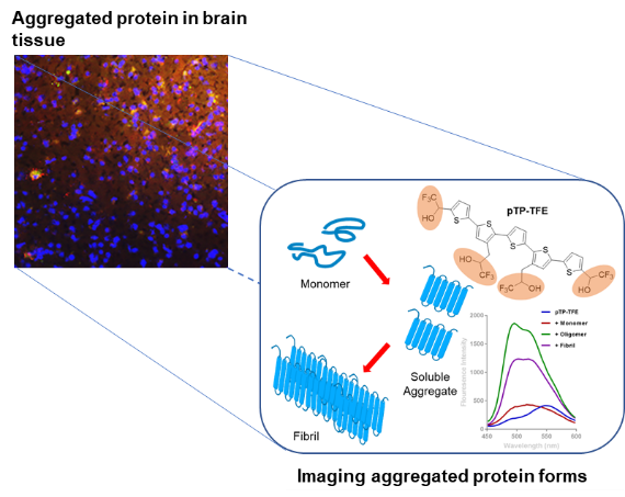
i) The development and characterisation of novel imaging probes that are specific and selective to biological markers and are sensitive to biochemical changes associated with disease mechanisms. For this a particular research interest is imaging probes for imaging neuroinflammation and misfolded aggregated protein associated with neurodegenerative disorders, as seen here:

A fluorescent molecular imaging probe with selectivity for soluble tau aggregated protein. Zhao Y et al., Chem. Sci. 2020. 11: p. 4773-4778
ii) The development of novel radiosynthetic methods for radiolabelling with short-lived PET radioisotopes such as carbon-11 (t½ = 20 min ) and fluorine-18 (t½ = 110min). The primary objective of this research is to develop rapid efficient radiosynthetic processes with an economy of reagents and few reaction steps to be able to produce radiopharmaceuticals on a routine and reliable basis for PET research studies and/or diagnostic services. In addition, by increasing the range of radiolabelling reagents and methods, we aim to widen the scope of compounds that can be developed for application as imaging probes, as demonstrated in this example:

CuI-Catalyzed 11C Carboxylation of Boronic Acid Esters: A Rapid and Convenient Entry to 11C-Labeled Carboxylic Acids, Esters, and Amides. Riss, P.J. et al, Angewandte Chemie-International Edition, 2012. 51(11): p. 2698-2702.
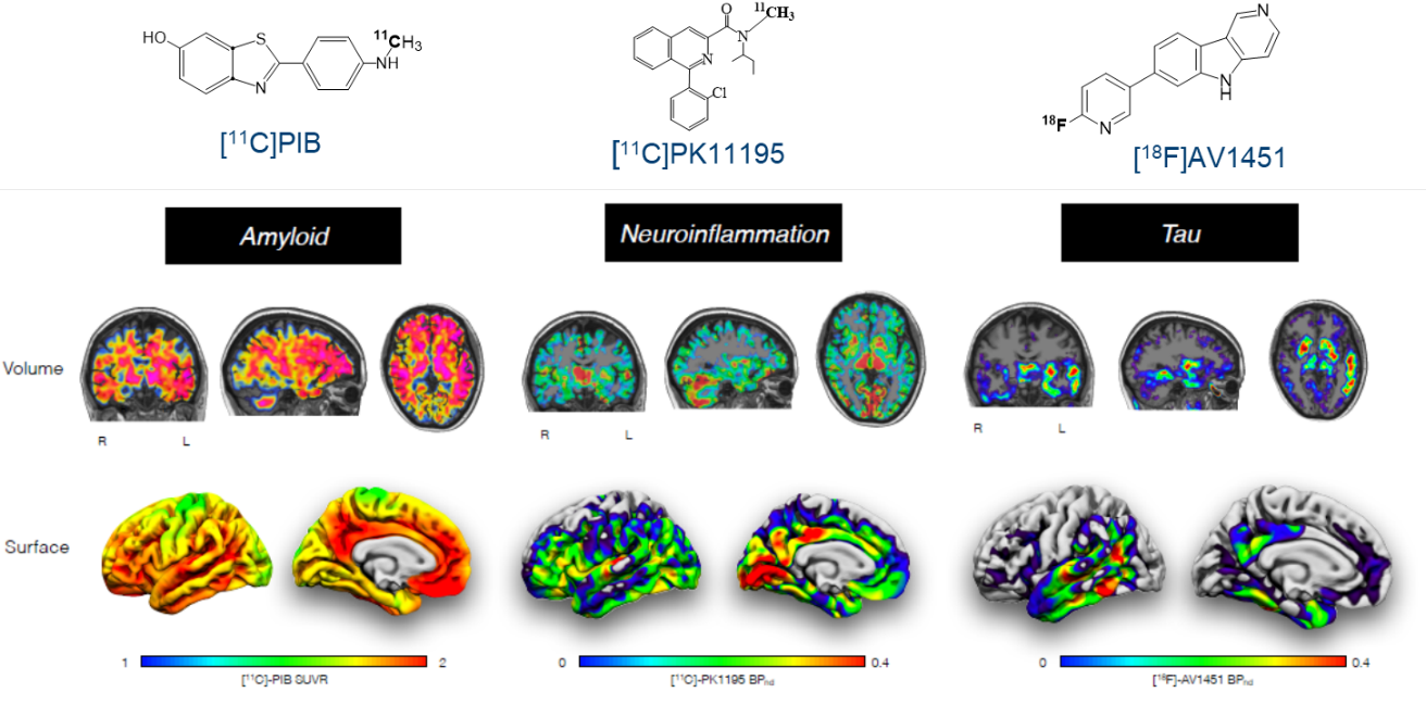
iii) Through collaboration with a wide range of biomedical research groups based at the School of Clinical Medicine I undertake preclinical and experimental medicine imaging research. This consists of applying the novel PET probes to investigate mechanism of human diseases, with a focus on neurological and mental disorders. In addition, we aim to apply the imaging techniques towards earlier detection and improved diagnosis of diseases and create a platform for the development of novel drugs and treatment evaluation, as below:

Delineation by PET imaging of the various pathological components of neurodegenerative disorders
F-18-AV-1451 positron emission tomography in Alzheimer's disease and progressive supranuclear palsy. Passamonti, L., et al,. Brain, 2017. 140: p. 781-791.
Neuroimaging of Inflammation in Memory and Related Other Disorders (NIMROD) study protocol: a deep phenotyping cohort study of the role of brain inflammation in dementia, depression and other neurological illnesses. Bevan-Jones, W.R., et al, BMJ Open, 2017. 7(1)
Neuroinflammatory and morphological changes in late-life depression: the NIMROD study. Su, L., et al, British Journal of Psychiatry, 2016. 209(6): p. 525-526.

