New microscopy method sees how protein forms clusters in living cells
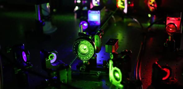
As part of the new method, the researchers used so-called HILO (highly inclined and laminated optical sheet) microscopy. Image courtesy of Thorsten Hugel, CIBSS, University of Freiburg
Using a newly developed microscopy method, researchers from Cambridge and Freiberg have been able to see for the first time how protein clusters form in living cells.
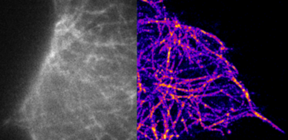
Super resolution microscopy may lead to better understanding of Alzheimer’s disease
Images of soluble tau using conventional microscopy (left) and super resolution microscopy (right), courtesy Klenerman Lab
Researchers have been able to observe and measure tau aggregates replicating in cells for the first time, in a key process that underpins the development of Alzheimer’s disease.
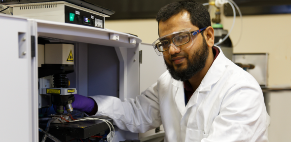
When a potentially useful molecule is first synthesised, it is often produced in quantities no larger than a pinch of salt. Oliver Griffiths has been scaling up promising reactions so that they are more useful for industrial applications.
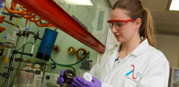
Linking drugs together can be more effective at inhibiting protein function than a single drug. Radu Costin Bizga Nicolescu investigated how drugs can combine to be more effective against diseases such as cancer.
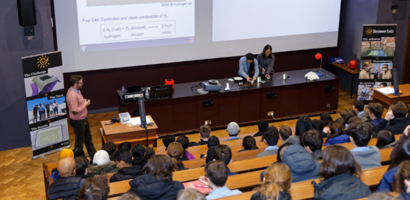
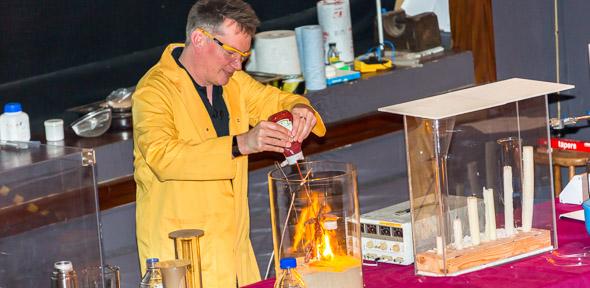
Alongside inventing new ways to create sustainable fuels for a circular economy, members of the Reisner group are telling the world about their results through outreach.
Nick Cowan (Downing College 1969) won our Chem@Cam cryptic crossword competition, and has received a colourful and sustainable Chemistry Department travel cup.
“Empowering future generations through education is at the core of everything we do,” says David Izuogu, who founded the Africa of Our Dream initiative in 2018 while completing his PhD in theoretical chemistry here.
Please join us in celebrating Dr Luke Abraham’s new title of Director of Research, which is a top rank on the Senior Researcher pathway and is considered to be equivalent to Professor (Grade 12).