Spectrally-resolved single-molecule fluorescence microscopy
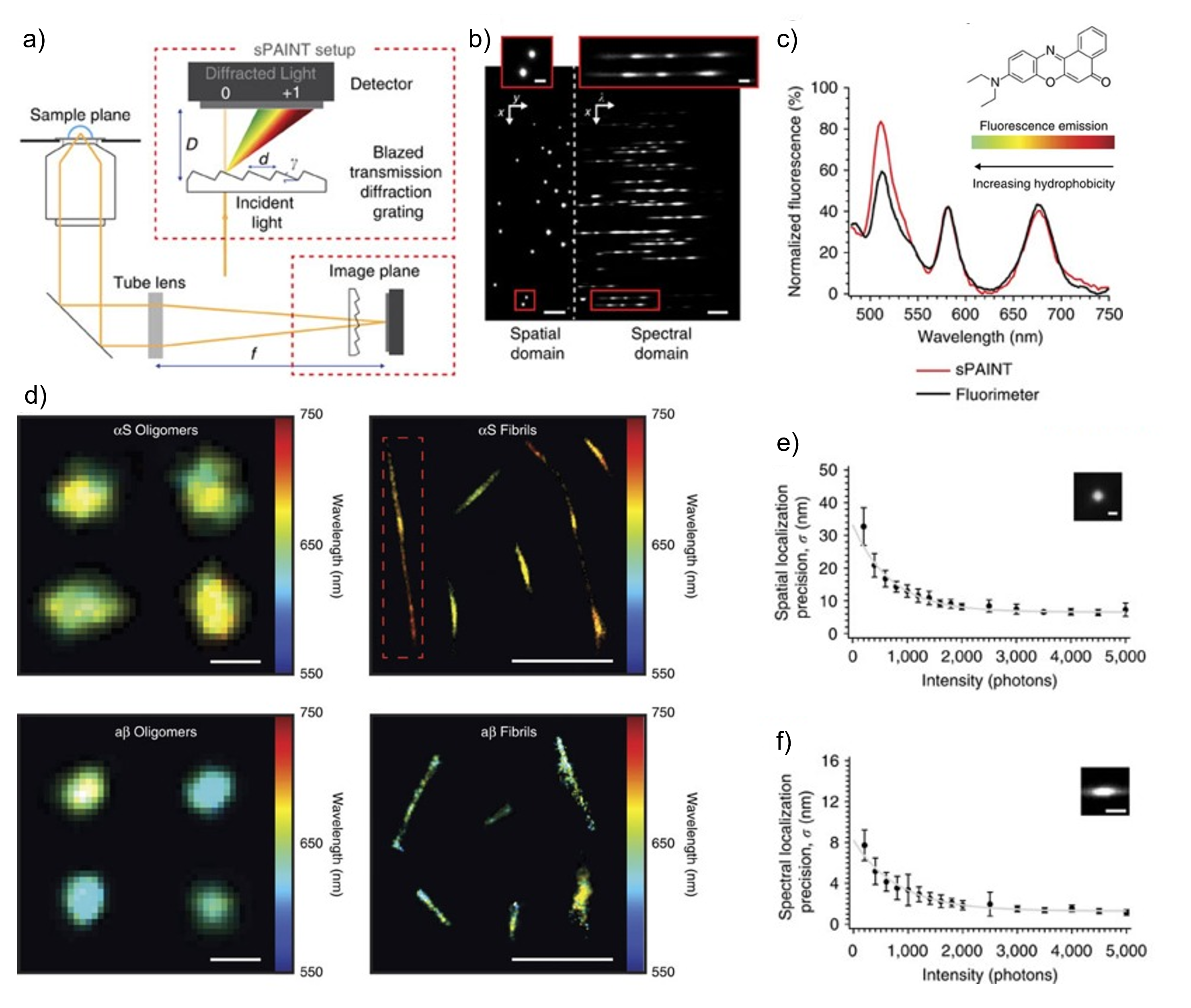
The Lee lab developed spectrally-resolved PAINT (points accumulation for imaging in nanoscale topography) or sPAINT, which simultaneously records the spatial position and emission spectrum of single dye molecules to super-resolve an image. For example, the fluorescence emission wavelength of the phenoxazone-based dye nile red (NR) is known to be sensitive to the hydrophobicity of its environment, and we have exploited this with sPAINT to super-resolve biological structures in the hydrophobicity domain. sPAINT is easily implemented by inserting a transmission diffraction grating into the optical path of a localization-based super-resolution microscope, enabling all the information to be extracted simultaneously from a single image plane.

Principles of the spectral-PAINT technique. a) Optical set-up for sPAINT showing the use of a diffraction grating to obtain spectral information and b) a typical image of nile red data. c) Coparison of fluorescent spectrum recorded by sPAINT and a fluorimeter. d) Representative sPAINT hydrophobicity images of oligomers and fibrials using the dye nile red (scale bar is 100 nm). e) Locialisation precision and f) spectral precision as a function of detected photons per molecule.
