Molecular origins of adaptive immunity
Using super-resolution imaging to study adaptive immunity
Cells constantly interact with their environment, using surface receptors to interact with and bind external stimuli, forming a signalling complex which leads to a cell response. An especially important example occurs in the adaptive immune system where T cells make close contacts with antigen presenting cells (APCs) while scanning for potential threats. Extraordinarily, in order to be effective, the immune system demands that a response is powerful (to neutralise the threat) and also highly specific, to avoid auto-immune attack or disease. While the role of T and B cells within the wider immune system is known, the interactions of these cells with external stimuli and the molecular processes that lead to an adaptive immune response (triggering) are areas of significant debate.
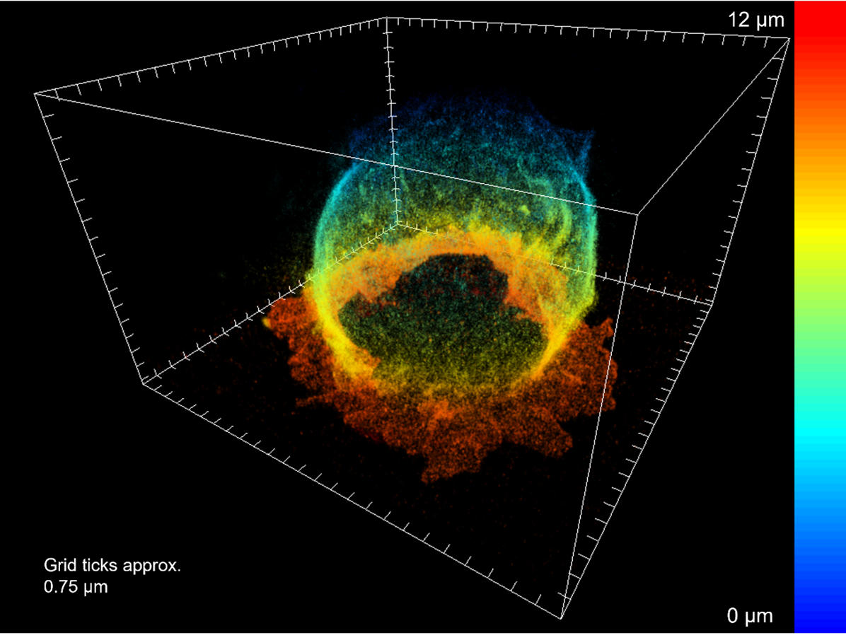
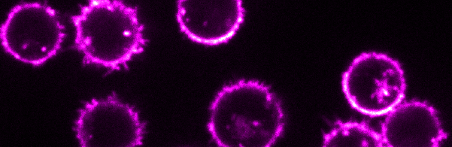
Figure. A Jurkat T cell interacting with a PLL coated surface. The basal surface of the T cell typically interacts using membranous protrusions called microvilli and then spreads out across the opposing surface in its search for an antigen.
As part of a long-running collaboration with both Professor Sir David Klenerman’s lab (University of Cambridge) and Professor Simon Davis’ Lab (University of Oxford) we use advanced imaging techniques such as 3D super resolution microscopy to better understand the resting T cell, and how changes in protein distribution or the cell morphology effects signalling outcomes.