Current Research Themes
Three-dimensional (3D) super-resolution (SR) and single-molecule localisation microscopy (SMLM) enables observation of the native morphology of complex biological structures by determining the 3D position of single fluorescent molecules bound to a target of interest. From imaging at different focal planes and interferometry to point spread function (PSF) engineering, TheLeeLab is at the forefront of the development of new 3D-SMLM methodologies, and houses several bespoke 3D microscopes, including two double-helix (DH-)PSF platforms and a single-molecule light field mircoscope.
Early Stages of Adaptive Immunity
The DHPSF is a 3D technique that transforms the 2D PSF of a traditional wide-field microscope into two gaussian intensity distributions (lobes) whereby the mid-point of the two lobes encodes lateral (xy) position and the angle between them encodes axial (z) position over 4 μm. The DHPSF is the current state-of-the-art 3D-SMLM technique, and we use it to image immune cells (T and B Cells) to study the early stages adaptive immunity. Figure 1 shows the membrane of a fixed T Cell imaged using the DHPSF.
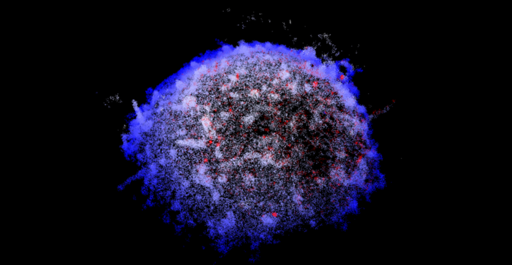
Figure 1: Apical T cell membrane (white) and CD45 (red) imaged in 3D
captured using the double helix point spread function (DHPSF).
Single Molecule Light Field Microscopy
In 2020, TheLeeLab introduced a new type of 3D-SMLM methodology called single molecule light field microscopy (SMLFM) in collaboration with Cambridge Advanced Imaging Centre (CAIC). SMLFM places a refractive microlens array (MLA) in the back focal plane (BFP) of a traditional wide-field microscope to obtain the 3D position of point emitters through disparity (parallax). Research in TheLeeLab continues to explore and extend the capabilities of SMLFM for applications in single particle tracking and systems with a low signal-to-noise ratio. Figure 2 shows some 3D super-resloved immune cells captured via SMLFM.
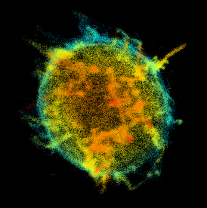
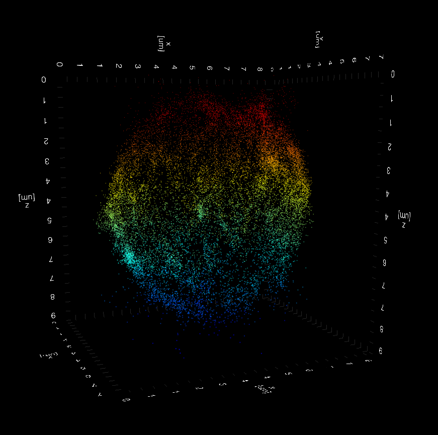
Figure 2: Apical surface of a T cell membrane (left) and the whole B cell dristribution of the B Cell Receptor (BCR, right)
captured using single molecule light field microscopy (SMLFM).
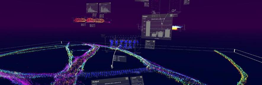
vLUME: 3D Virtual Reality for 3D-SMLM Analysis
A collaboration between TheLeeLab and LUME VR saw the development of a software package based on the visualisation and subsequent analysis of 3D-SMLM data using virtual reality. vLUME enables the user to analyse their 3D-SMLM data through immersion with VR using Oculus Rift and was published in Nature Methods in 2020. Our collaboration with LUME continues to explore alternative ways of visualising (and analysing) complex 3D-SMLM data.